
Advanced Imaging at OAESH
Ottawa Animal Emergency & Specialty Hospital | Specialty | Advanced Imaging
At Ottawa Animal Emergency & Specialty Hospital (OAESH), our team uses state-of-the-art imaging services to provide accurate, timely diagnoses for pets with a wide range of health concerns. Our advanced diagnostic tools allow veterinarians to identify and monitor diseases quickly, supporting precise treatment plans that improve your pet’s quality of life.
Our facility offers:
- Digital radiographic imaging (X-ray)
- Ultrasound imaging (echography)
- CT-Scan (computed tomography)
Whether for emergency evaluation or ongoing monitoring, OAESH ensures your pet receives thorough, compassionate, and expert care.
Digital Radiographic Imaging (X-Ray)
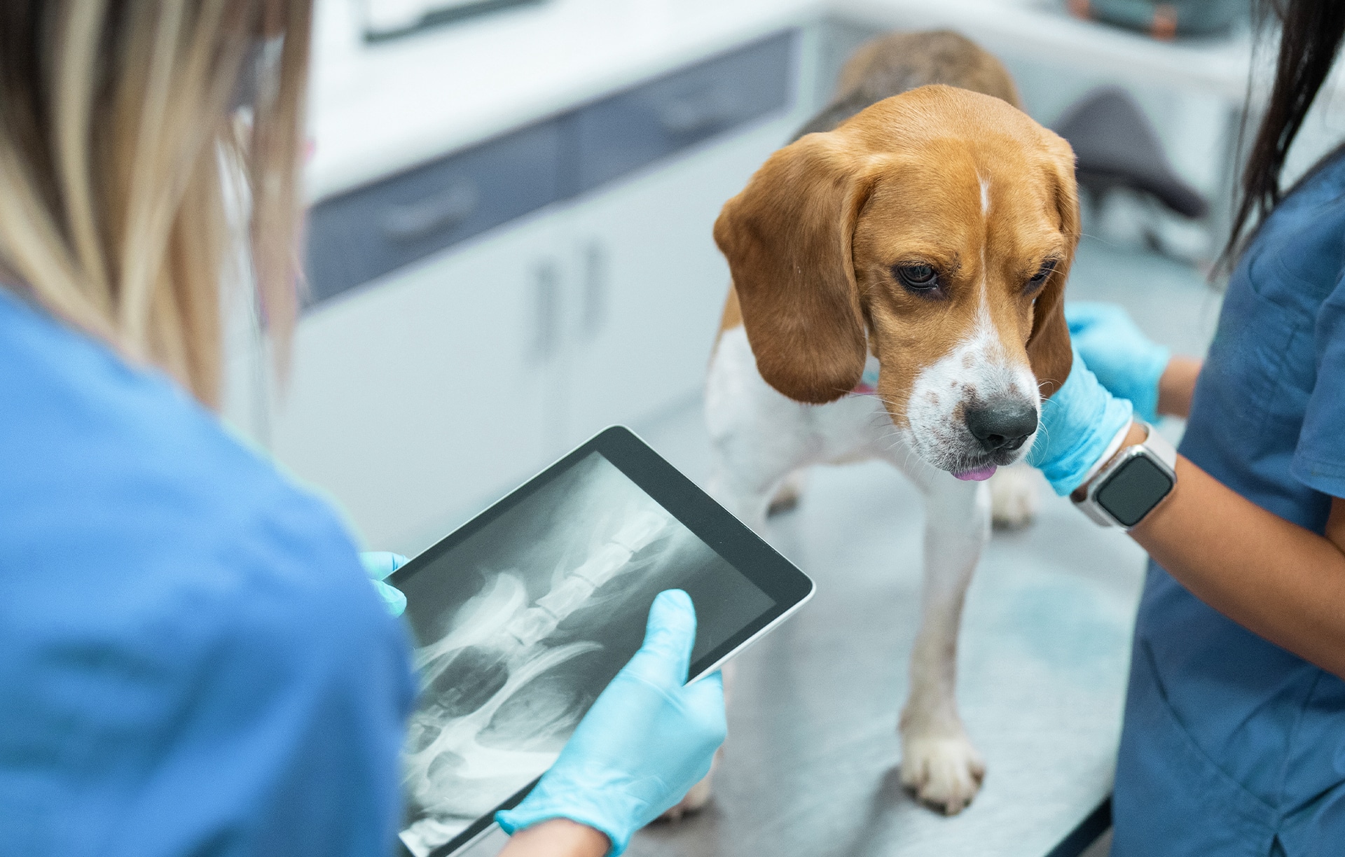
Digital radiography is an essential tool for diagnosing many conditions in pets. Our X-ray services include:
- Upper and lower limb radiographs for bone, intervertebral disc, or joint lesions
- Neck radiographs for upper respiratory tract conditions, including tracheal, laryngeal, or esophageal injuries
- Thoracic radiographs (3 views) for lungs, heart, and blood vessels
- Abdominal radiographs (3 views) for stomach, intestines, liver, spleen, kidneys, bladder, and tumors
Our digital radiography system produces highly detailed images quickly. Images can be computer-enhanced for optimal diagnostic clarity and saved to CD-ROM or USB for your records. All emergency radiographs are reviewed by board-certified ACVR radiologists, and an official report is included with your pet’s medical record.
Ultrasound Imaging (Echography)
Our veterinary ultrasound allows precise, real-time evaluation of thoracic and abdominal organs, helping detect fluid accumulation, masses, or other abnormalities. Ultrasound is frequently used to:
- Assess abdominal organs and thoracic structures
- Perform fine-needle aspirates of masses or lesions for diagnostic purposes
- Obtain sterile urine samples for bacterial culture in suspected urinary infections
Most ultrasound procedures require mild sedation to ensure accurate assessment and safe sampling. Ultrasound provides a non-invasive, highly informative method to support diagnosis and treatment planning.
CT-Scan (Computed Tomography)
CT scanning is performed under general anesthesia or heavy sedation and offers high-resolution imaging of complex structures. CT scans are particularly useful for:
- Evaluating complex fractures and vertebral disc herniation
- Diagnosing nasal cavity or chronic ear conditions
- Pre-surgical evaluation of masses
- Whole-body imaging for cancer staging
CT allows our specialists to visualize intricate anatomy that may not be captured with X-ray or ultrasound, aiding in accurate diagnoses and treatment planning.
To send a referral, please fill out this form and send it to [email protected], or visit out referrals page!